Category Archives: Biology

Posts related to my Biology A Level

Endocytosis & Exocytosis

Bulky substances are transported into and out of the cell by endocytosis and exocytosis.

Endocytosis:

This is the infolding of the cell membrane to form vesicles. A portion of the membrane invaginates to envelope the contents and draw them into the cell. Once inside, the vesicles are known as intracellular vesicles.

There are 3 types of endo-:

1. Phagocytosis (cell eating)

When a bacterium or other solid item is engulfed by a cell membrane, the intracellular vesicle that was hence formed comes into contact with a lysosome. The lysosome fuses its membrane with the vesicle to release its catabolic enzymes which break down the solid.

2. Pinocytosis (cell drinking)

This occurs when a liquid is engulfed by a cell. In cells with multiple microvilli, such as the intestinal epithelial cells, there are pinocytosis channels between the microvilli which are constantly budding off vesicles of liquid. The cell membrane wraps around a fluid and pinches off, drawing in the liquid in a vesicle, the contents of which are then either broken down or absorbed into the cytosol.

3. Receptor Mediated Endocytosis.

This is a very specific type of pinocytosis because it involves receptors….

Falling asleep now, so I’ll finish this off tomorrow. 🙂

Leave a comment

Filed under Biology

The Plasma Membrane

An intact membrane is essential to a cell. If the plasma membrane is disrupted, the cell loses its content and dies. The membrane is very important; two vital functions are:

The regulation of the entrance and exit of molecules: The interior and exterior of the cell is mainly fluid. The membrane functions to keep the intracellular fluid constant despite molecules such as nutrients and waste constantly moving in and out.

Communication: The components of a membrane signal other cells as to what type of cell it is. It may also serve as receptors for various signal molecules that affect the cell’s metabolism.

Membrane Models:

At the beginning of the last century, scientist noted that lipid-soluble molecules entered the cells more rapidly than water-soluble molecules. This caused them to think that lipids were a component of the plasma membrane.

Later it was discovered that it consists of phospholipids and proteins. Phospholipids are lipids in which one of the fatty acid groups is replaced by H3PO4. The phosphoric acid is hydrophillic, the rest of the molecule is hydrophobic.

The ‘Fluid’ Membrane

A membrane is held together by weak hydrophobic interactions. Most membrane lipids are able to drift laterally within the membrane and occasionally flip vertically, known as ‘flip-flopping’. Phospholipids move quickly along the membrane plane, where as the proteins move relatively slowly.

Unsaturated hydrocarbon tails enhance membrane fluidity because the kinks at the carbon-carbon double bonds hinder close packing of the phospholipids. Membranes solidify at the critical temperature. This is lower in a membrane with a higher concentration of C=C bonds.

Cholesterol found in the plasma membranes of eukaryotes modulates membrane fluidity by keeping the membrane fluid in cold environments and solid in hot temperatures. Cells may also the concentration of unsaturated fats to better suit their environment.

Integral proteins, which are inserted into the membrane have hydrophobic regions, surrounded by the hydrophobic areas of the phhospholipids. Their hydrophillic ends are exposed at both sides of the membrane.

The proteins in the plasma membrane may provide a variety of major cell functions:

  • Transport
  • Intercellular joining
  • Enzymatic activity
  • Cell-cell recognition
  • Signal transduction
  • Attachment to the cytoskeleton and extracellular fliud.

Substances can be moved through the membrane via: Active Transport; Diffusion; and Osmosis.

Active Transport is the movement of molecules from an area of low solute concentration to an area of high solute concentration, against the concentration gradient, in a process that requires energy.

Diffusion is the passive movement of molecules from an area of high concentration to an area of low concentration.

Osmosis is the movement of water molecules from a region of high water concentration to a region of low water concentration through a semi-permeable membrane.

Diffusion is influenced by:

  • The permeability of the membrane
  • The shape and size of the molecule to be transported
  • Number of proteins on the cell surface
  • Concentration of molecules on either side of the membrane
  • Surface area of the membrane

Facilitated diffusion is a method of diffusion that uses proteins to transport substances that find it difficult to pass through a membrane. e.g. polar molecules. The proteins are known as carrier proteins.

Fick’s Law

Rate of diffusion is proportional to the surface area multiplied by the difference in concentration, all divided by the thickness of the membrane.


Rate of diffusion (fish)  Surface area x Concentration gradient

thickness of membrane


Osmosis – Thermodynamics

1 molecule of water will move quickly if heat is applied, or if the water concentration in a solution is high.

If water molecules are moving from left to right, then the potential energy is greater on the left than on the right. The potential energy is known, in Biology, as water potential.

Water diffuses from an area of high water potential to a region of low water potential through a semi permeable membrane. Water potential can be regarded as the tendency of water to leave a solution.

If solute molecules are present, they always slow down the movement of the water molecules in a solution. The tendency of the water to leave the solution is reduced because water is always attracted to the solute.

Water Potential Gradient

High          Pure water = 0 kPa


Dilute solution = -500 kPa


Low            Concentrated solution = -1000 kPa

Water potential is never positive. When the potential is more negative, water will flow into the cell.

Isotonic – two solutions are of the same water concentration, and as such there is no net movement of water.

Hypotonic – The water potential outside of the cell is greater than the intracellular potential. As such, there is a net inflow of water. The inside of the cell is more negative.

Hypertonic – The water potential inside of the cell is greater then the extracellular potential. As such, there is a net outflow of water. There inside of the cell is less negative.


Active Transport

Active transport requires energy in the form of ATP. it trasports molecules and ions in a direction that is not natural to the normal flow. This means that there will be many mitochondria present.

The following use ATP to transport molecules and ions:

1. Membrane pumps

  • An active transport mechanism that moves ions in order to obtain polarisation
  • For active transport two factors need to be considered: concentration and electrical charge.
  • Ions generally diffuse to form an area of high concentration to an area of low concentration and are attracted to regions with an opposite charge. Therefore we take into consideration both the concentration and elecrtochemical gradient.
  • Cells maintain a potential difference across the membrane. Many studies have shown that the inside of a cell is -ve and therefore cations are attracted and anions repulsed.
  • however, their relative concentrations inside and outside the of the cells helps to decide which way they move.
  • Three common ions to be transported are K+, Na+ and Cl-

1. Sodium Potassium pump

  • Cell surface membranes have pumps that are intrinsic proteins that span the membrane. The sodium pump removes Na+ from the cell. K+ is taken into the cell and so is coupled with the Na+ pump. It is therefore known as the Na+/P+ pump.
  • The pump requires more than one third of the ATP produced by a resting animal. It is very important.
  • The pump is essential for:
  1. controlling cell volume (osmoregulation)
  2. Maintaining electrical activity in nerve and muscle cells
  3. Driving active transport of other substances (e.g. sugars and amino acids.)

Active transport in the intestine:

Soon after feeding there is a high concentration of food in the gut. Absorption is mainly due to diffusion but it is very slow and so it is coupled with the active transport and the movement of Na+. As the sodium is actively transported out by the Na+/K+ pump, it will start to diffuse back in. A membrane rquires both Na+ and glucose and so another pump is used that transports glucose at the same time as Na+.

Leave a comment

Filed under Biology

The Cell Membrane

So as it stands, I’m still trying to deal with the backlog of work from September, but I’m almost there, not much left to catch up on now. Anyway, this post is about the cell membrane and the history of its discovery.

1925 – Gorter and Grendel measured the amount of phospholipid extracted from red blood cells and determined that there was just enough to form a bilayer around the cell.

1935 – Danielli and Davson suggest that globular proteins are part of the membrane and proposed to sandwich model.

Late 1950s – Electron microscopy had advanced and J.D. Robertson suggested that all membranes in various cells have basically the same composition, leading to the unit membrane model.

1972 – J. S. Singer and G. L. Nicolson suggested a new structure for the cell membrane, the “Fluid Mosaic Model”. They proposed in part that the membrane is a phospholipid bilayer in which protein molecules are either partially or wholly embedded. This structure is still widely accepted at this time.

Leave a comment

Filed under Biology

Cells

So this was the topic that kicked off my AS course, you do a lot about cells in GCSE but this just goes into a bit more detail, so it’s not that much harder. Make sure to learn all the key definitions though!
Light Microscopy

  • Light microscopy is the oldest and most widely used form of microscopy
  • Specimens are illuminated with light, which is focused using glass lenses and viewed using the eye or photographic cells.
  • Specimens can be dead or alive, and often need to be stained to me made visible. Iodine, which stains starch blue/black, is used for plant cells, and methylene blue used for animal cells.

A couple of important definitions:

Magnification – refers to the microscope’s power to increase an object’s apparent size.

Resolution – refers to the microscope’s power to show detail clearly, so that the use is able to distinguish two points close together.

The resolving power of a microscope is limited by the wavelength of light (400-600 nm). If objects in the specimen are smaller than the wavelength of radiation being used, they do not interrupt the waves and so are not detected.

Preparation of Slides

Slides need to be made so that the specimen can be viewed time after time for a number of years.

  1. Fixation – Chemicals are used to preserve the specimen so that they are not distorted over time.
  2. Dehydration – Water is removed from the specimen using a solvent, generally ethanol. This is because water molecules with deflect the beam of electrons away from the specimen.
  3. Embedding – Specimens are put into wax/resin to hold the structures in place so that thin slices can be made.
  4. Staining – Most biological material is transparent. Therefore different stains are used to highlight different organelles.
  5. Mounting – Mounting on a slide will protect the specimen so that it is suitable for viewing over a long period of time. A coverslip is generally placed over the specimen.

Eukaryotes:

These are cells that contain a double-membrane bound nucleus and other membrane bound organelles, such as mitochondria, endoplasmic reticulum and the golgi apparatus.

Nucleus – This is the most visible structure in a non-dividing cell, and contains most of the cell’s genetic material. The membrane bound area that surrounds the nucleus is known as the nuclear envelope. The nuclear envelope is a double-membrane, part of the endomembrane system, consisting of an inner and outer membrane, seperated by a gap of 20-40 nm. The two membranes fuse at the tips of the nuclear pores, which allow ribosomes and RNA out of the nucleus. Within each nucleus is one (or more) nucleolus, which is an area of concentrated DNA, organised by histones.

The nucleolus is roughly spherical and functions in the synthesis of ribosomes. It consists of nucleolar organisers (specialised chromosomes) with multiple copies of the genes for ribosome synthesis. They will have considerable amounts of rRNA and protein, representing ribosomes in various stages of construction. Generally, only one nucleolus is present, however there may more more than one, dependent on the cell species and the stage of cell cycle.

Ribosomes – These are essential for the process of protein synthesis. They are built up of two subunits; a large one with two tRNA binding sites, and a smaller one which associates with mRNA in a binding groove. They are the smallest eukaryotic organelles, and they do not have a membrane. Free ribosomes are located within the cytosol and produce the proteins needed in the cell. Bound ribosomes are attached to the ER; these produce the proteins needed for secretion.

Endoplasmic Reticulum – This a network of flattened sacs, called cisternae, and membranes throughout the cell. The membrane is continuous with that of the nucleus. There are two parts the the ER: the smooth endoplasmic reticulum and the rough endoplasmic reticulum. The SER is involved in synthesising lipids and steroids, and also carbohydrate metabolism. It also plays a part in the detoxification of drugs and poisons (e.g. alcohol). It is called “smooth” as there are no ribosomes bound to it. The RER has a rough surface, due to the ribosomes bound to the cytosolic side. As the ribosomes feed polypeptide chains into the RER’s lumen, it folds them into a functional proteins. Often carbohydrates are attached to form glycoproteins. Once the ribosome has finished feeding in the polypeptide, it detaches from the ER and moves back through the cytosol.

Golgi Apparatus – Anything made the ER is transported here in vesicles, where it is modified and then sent on to another destination. The golgi, similar to the ER, is made up of a series of folded cisternae, which have a cis face, and a trans face. The cis face is the receiving side of the golgi, and this is where transport vesicles from the ER attach to the apparatus. The trans face is the shipping face; here, vesicles bud off to carry molecules to other destinations, often to be secreted out of the cell. Secretory cells, such as those in the pancreas, have larger, more prevalent golgi.

Lysosomes – These are hydrolytic enzyme complexes surrounded by a single wall membrane. They bud off from the golgi, which produces the enzymes used to break down macromolecules. The lysosomes are capable of maintaining a high concentration of H+ ions by actively pumping them into the lysosymal lumen. Due to the anabolic nature of the enzymes, excessive leakage of the lysosome can result in autodigestion. It is protected from self digestion by the inner surface of its membrane. Lysosomes play a major part in cytosis.

Prokaryotes


Prokaryotes are the most primitive cell, appearing on Earth ~3.5 billion years ago as the first sign of life. They have no membrane mound organelles, most particularly, they have no double-membrane bound nucleus. They still contain DNA, however their genetic material is shaped in a circular plasmid, rather the the eukaryotic double helix, these strands are left lying free in the cytosol. The ribosomes in prokaryotes are a lot smaller than those in eukaryotes, but the cells do have a similar metabolism. Generally they are much smaller in size, typically between 0.5-10 microns in diameter. In bacteria is the only place that nitrogen fixation occurs. Nitrogen is absorbed from the atmosphere and converted into nitrates by the organisms.

Leave a comment

Filed under Biology

Proteins

Moving on from carbohydrates, we come to proteins. Proteins are another macromolecule, also known as polypeptides, and are formed when amino acids are linked into long chains via peptide bonds. There are many different types of proteins important to life such as enzymes and the proteins in the fluid plasma membrane of a cell.

There are seven different classifications of proteins:

Enzymes Biological catalysts that speed up chemical reactions in the body. E.g. ATPase, an enzyme found in all cells which catalyses the breakdown and formation of ATP.
Structural proteins Proteins that form part of a body of an organism. Keratin, a major component of hair is a structural protein.
Signal proteins These carry messages around the body. For example, the hormone insulin, which is involved in the controlling of blood glucose levels.
Contractile proteins These are proteins involved in movement, such as actin and myosin, which allow muscles to contract.
Storage proteins These are proteins that store nutrients, for example the albumen in eggs.
Defensive proteins Proteins that form part of the immune system, mainly the blood antibodies.
Transport proteins These transport materials around organism, e.g. the haemoglobin in blood.

Proteins are made up of combinations of several different amino acids; there are 20 amino acids in total. Amino acids are composed of an Amine group (NH2), a Carboxylic acid group (COOH) and an R group. The R group is replaced with any of the 20 R side chains, which determines the properties of the amino acid formed. The simplest amino acid is glycine, which has an R group of just a single hydrogen atom. The amino group, carboxylic group, a hydrogen atom and the R group are each bonded to a central alpha carbon atom with a covalent bond. Some R groups are basic, others alkali, some hydrophilic, others hydrophobic, and some contain the compound benzene. Amino acids are amphoteric, which means they can act as acids or alkalis.

An amino acid can also be known as a monopeptide. Through condensation polymerisation, two or more amino acids can combine to form a dipeptide, or a polypeptide. The bonds that form between each amino acid are called peptide bonds. When a polypeptide consists of enough amino acids, it is known as a protein.

Proteins are each built up of four structures, each structural layer determining the next. The primary structure is the exact sequence of the amino acids that make up its polypeptide chain. By switching the position of just two of these amino acids the whole protein structure could be changed.

A protein’s secondary structure is the way in which the polypeptide chain coils or folds into either an alpha-helix or a beta-pleated sheet. The shape of these structures is held b regularly spaced hydrogen bonds which form between the N-H group of one amino acid and the C=O group of another.

The tertiary structure is the 3D shape of the polypeptide chain; proteins can be classified as either fibrous or globular, depending on their tertiary structure. Fibrous proteins are composed of parallel polypeptide chains cross linked to form long fibres or sheets. They are usually insoluble in water and physically tough, which makes them suitable for their mainly structural functions. In globular proteins, the chain of amino acids is tightly folded to form a spherical shape. Many of them are folded so that their hydrophobic ends are inside the molecule, and the hydrophilic ends facing outside, making the molecule water soluble. Many hormones, antibodies and enzymes are globular proteins. The precise shape of the globular protein determines its every property.

If the bonds holding a protein together are broken, then denaturation occurs. The polypeptide is chemically unchanged but the tertiary structure of the chain is lost, and hence they lose their specific shape and cannot function. The process of denaturation is nearly always irreversible, and can be caused by changes in pH, temperature or salt concentration.

The quaternary structure is the way in which more than one polypeptide chains bond together, and the way in which they are arranged.


2 Comments

Filed under Biology

Carbohydrates

Continuing on molecules that are important to life, carbohydrates are a huge part of the syllabus which I covered. A comparison of their composition and structure is a common essay title and one which I suggest is learnt. Knowing how to draw the sugar monomers, and also the glycosidic bonds between them, is another important skill.


Carbohydrates:

All carbohydrates contain carbon, hydrogen and oxygen and have the common formula Cx(H2O)y. Carbohydrates include compounds such as sugars, starch, glycogen and cellulose.

Monosaccharides

Simple sugars all have the formula (CH2O)n, meaning that the three component elements are always present in the same ratio. Monosaccharides are grouped according to the value of n, which can range from 3-7.

n Sugar
3 Triose
4 Tetrose
5 Pentose
6 Hexose
7 Heptose


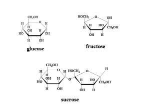
Glucose is the most common hexose, and has the formula C6H12O6. However, this formula does not show how the atoms are structurally arranged, and glucose can have a number of different shapes. Isomerism is when the same atoms are arranged differently to form many shapes. Each compound with the same chemical formula, but different structural formula is called an isomer.

Glucose can form a straight molecule, or two different isomeric rings; alpha glucose and beta glucose. The only difference between the two rings is the position of the hydrogen and hydroxyl groups on carbon atom 1. When the hydrogen atom is above the carbon atom, the ring is called alpha glucose. Beta glucose has the hydroxyl group above the carbon atom.

These different arrangements have very different properties. Alpha glucose molecules combine to form starch, where as beta glucose molecules form cellulose.

Disaccharides

Two monosaccharides can join together to form a disaccharide in a condensation reaction. Different pairs of simple sugars form different disaccharides. The bond formed between the two monosaccharides that joins them is called a glycosidic bond.

Monosaccharides Disaccharide formed
Glucose + Glucose Maltose
Glucose + Fructose Sucrose
Glucose + Galactose Lactose


Condensation involves the removal of water, so ion the process of joining two sugars, one water molecule is formed. The opposite of condensation is hydrolysis, which breaks down the disaccharide into its component part by the addition of water.


Long chains of many monosaccharides joined together can be formed in the same way. A molecule formed of several simple sugars is called a polysaccharide. The process of making polysaccharides is called polymerisation. Polysaccharides are polymers, large molecules formed of multiple repeating units, called monomers. They have the generic formula (C6H10O5)n. Polysaccharides are not particularly soluble in water, are not sweet, and do not form crystals. The properties of polysaccharides are determined by the number and type of the monomers joined together, and also the shape that they form. Polysaccharides can be straight, helical, coiled or branched.

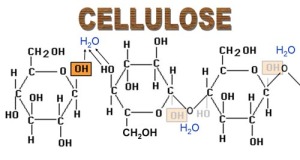
Each carbon atom in a monosaccharide unit is given a number, (in glucose from 1-6), different carbon atoms bond in different ways to form different chains of polysaccharides. The reaction between the hydroxyl groups at carbon atom 1 of one molecule, and atom 4 of another molecule forms a 1-4 glycosidic bond, many of which form long, straight chains. Branched chains have one or more 1-6 glycosidic bonds, which form between the hydroxyl groups on a carbon atom 1 and a carbon atom 6.


Starch is formed of hundreds of repeating alpha glucose molecules. It is compact due to its tight globular structure. Its natural spiral structure is compressed due to the attraction between the different molecules. When an Iodine molecule is trapped in the spiral of the starch, it changes colour from brown to blue/black, which is what we see when testing for starch in food tests. In plants, starch is stored in the membranous organelles called plastids, e.g. chloroplasts. Starch is a source of organic carbon for making other substances and is also broken down during digestion into glucose for respiration.

Another storage polysaccharide is glycogen, it is found mainly in the liver and muscle cells. It has a similar roles and structure to starch, and since it is more abundant in animals, it is often referred to as ‘animal starch’. Glycogen is less dense and more soluble than starch as it has many more 1-6 glycosidic bonds. It can be broken down (hydrolysed) more readily than starch, as such animals have a higher metabolic rate than plants.

Cellulose is another polysaccharide, and it is a major component of plant cell walls. It is formed by beta glucose monomers linked by 1-4 glycosidic bonds. Cellulose is completely permeable, meaning that it allows water and other substances to pass through it into and out of the cell freely. Unlike starch and glycogen, cellulose cannot be hydrolysed easily. Animals such as cows are able to digest plant matter efficiently as their stomachs produce cellulase, an enzyme that speeds up the breakdown of cellulose. Since humans do not produce cellulase, we cannot obtain the nutrients contained in plant cells. Lingin is used by plants to strengthen cellulose further. It creates an impermeable lining on the xylem tubes, providing added support, and also helps prevent infection and decay.


Leave a comment

Filed under Biology

The Biological Importance of Water

The first topic that I covered in AS Level Biology was about molecules of biological importance.  Water is a substance that is in great abundance on this planet, and it holds some significant importance to our lives. Without it, we could not live, and not simply because we would die of thirst.  Some of the notes here are also relevant to AS Chemistry, but be careful, as different specifications might ask for information on different anomalous properties, so make sure you check your exam syllabus!

The Properties of Water


  • Water is a dipolar molecule:

A water molecule consists of two hydrogen atoms and an oxygen atom; however, the electrons in the covalent bonding are not shared equally. The oxygen atom has a greater electronegativity, meaning that it has a greater pull on the electrons. Due to this each water molecule has slightly negative and slightly positive regions.

  • Water molecules form hydrogen bonds:

The negative and positive ends of water molecules attract each other to form hydrogen bonds. These hydrogen bonds give water many of its unique properties. Compounds with molecules similar to the size of water are usually gases. Since each water molecule can form hydrogen bonds with up to 4 other water molecules, water is a liquid at room temperature.

  • Water is the universal solvent:

Polar and ionic substances have an electrostatic charge, so they are attracted to the charges on water molecules and dissolve readily. Non-polar substances, such as oil, do not dissolve in water, as they do not have charged molecules. When a salt dissolves in water, the ions separate and a layer of water molecules form around the ions. These layers prevent ions or polar molecules from clumping together, keeping the particles in solution.

  • Water has a high surface tension:

At an interface between air and water, a water molecule on the surface forms hydrogen bonds with other molecules around and below it, but not with air molecules above it. The unequal distribution of bonds produces a force called surface tension; this causes the water surface to contract and form a surprisingly tough film or ‘skin’.

  • Ice floats on water:

Water is at its most dense at 4oC. When water freezes the hydrogen bonds between the molecules forms a rigid lattice, that holds the molecules further apart then in liquid water. Ice, having expanded when freezing, is less dense than its liquid counterpart and so floats on water.

  • Water is adhesive and cohesive:

Water is ‘wet’ because it sticks to things. This is because its molecules can form hydrogen bonds with other polar substances. This is called adhesion. The attraction between molecules of similar substances is called cohesion. In this way water molecules stick together which allows water to enter and move along very narrow spaces, in a process called capillarity.

  • Important thermal properties:

Water has a high specific heat capacity meaning that it needs to gain a lot of energy to raise its temperature. Conversely it also needs to lose a lot of energy to lower its temperature. Water’s specific heat capacity is 4.2 kJ/g/oC

Water has a high latent heat of vaporisation which means a lot of energy is required to evaporate it. When it evaporates, water draws thermal energy out of the surface it’s on, which can be observed in sweating.

Water also has a high latent heat of fusion meaning that at 0oC water must lose a lot of thermal energy before it freezes, thus liquid water can reach temperatures of down to -10oC before it forms ice.

  • Other physical properties of water:

It is transparent to sunlight.

It has a relatively high density compared to air.

It is difficult to compress.

It conducts electricity (when it contains dissolved ions)

See if you can think of ways that these properties are important to life.

An important part of A Level Biology is being able to write a clear and concise essay on the topics you have covered, particularly as many exam boards set an essay question in each of their papers. Below is an example essay on the importance of water.

The Biological Importance of Water


Water has several unique properties that make it vital not only for human beings, but for all living organisms to survive. The most noticeable of its physical properties is that it is a liquid at room temperature, which is unusual for compounds with molecules of a similar atomic composition. This is due to the hydrogen bonds that form between each water molecule, and up to four others. Water being a liquid at room temperature provides a marine environment for organisms to live in, and also provides a liquid environment inside cells, which holds significant importance as metabolic reactions that are key to life take place in solution.

Water molecules are dipolar, meaning they have a positively charged and a negatively charged region. The charges of these areas attract polar and ionic substances that are dissolved in it, and the water molecules form a layer around each charged ion, keeping the substance in solution. Water is known as the ‘universal solvent’, this is because it dissolves much more substances than most common solvents.  This is of vital significance as all of the metabolic reactions essential for life take place in solution in the cytoplasm of living cells.

Another property caused by water molecules being dipolar is that water is adhesive, and this adhesion makes water stick to other polar substances, effectively making it ‘wet’. This allows water to move upwards through the very narrow xylem of tall plants, such as trees, against gravity. Continuous columns of water can also be pulled up to the top of trees due to its high tensile strength, meaning that water columns do not break easily. Also important to plants is water’s transparency. Water, being transparent and colourless transmits sunlight, enabling aquatic plants to photosynthesis, and also enabling us to see, as our eyes are coated in water.

There are also many thermal properties that make water so essential for life, for example its very high specific heat capacity, 4.2kJ/g/oC . This means that a lot of energy needs to be gained, or lost, in order to change the temperature of water, and so the environment inside organisms resists temperature changes that could cause it damage. Water also has a high latent heat of vaporisation which means mean that water needs a lot of energy to evaporate, and so draws this thermal energy from the surface it is on, cooling it as the water evaporates from it (this can be observed when we sweat to cool ourselves). Water’s high latent heat of fusion prevents the liquid environment of cells from freezing, and tearing the cells apart, as liquid water temperatures can drop to around -10oC before it begins to freeze.

Leave a comment

Filed under Biology, Chemistry